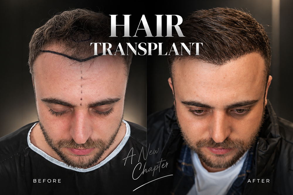
Hair transplant before and after galleries compress a full year of biology into two frames, which is exactly why they mislead. A result photographed at month 6 is not the result at month 12. This International Plus guide walks the timeline honestly: healing; the shed nobody warns patients about; the dormant stretch; the month the crown catches up. Photographs published by our clinic carry written patient consent and individual results vary.
Key Takeaways
- A credible hair transplant before and after comparison needs at least 4 time points since a single pair of frames hides the shedding phase entirely.
- Shock loss between weeks 2 and 6 is a normal telogen response, not graft failure because the follicle stays anchored while the shaft detaches.
- The crown reads later than the hairline, often by 6 months since whorl geometry scatters light across a curved surface.
- Photo protocol decides credibility: fixed distance, fixed lighting, dry hair, plus a donor area frame.
- Slow growth deserves a physician review at month 6, not a second surgery booked on impatience.
What Do Hair Transplant Before and After Results Show?
Hair transplant before and after results are a record of one surgical event followed by roughly 12 months of biological change. Photographs taken at day 0, month 6 and month 12 capture three different scalps. The final comparison reflects graft survival, hairline design, the growth cycle itself rather than surgical skill alone.
Photographs mislead when a reader treats them as a promise. A gallery shows a completed outcome without the donor supply behind the result. According to Fu and colleagues in Plastic and Reconstructive Surgery (2026), a multicentre study of frontal hairline correction found patient satisfaction driven by design decisions as much as by graft numbers. Every International Plus image is published with written patient consent, never as a guarantee. In practice, our team photographs each case at 4 review visits using International Plus protocols so progress is judged against a baseline rather than against memory. Candidacy for a hair transplant procedure is confirmed before any timeline discussion begins.
What Does the Month by Month Timeline Look Like?
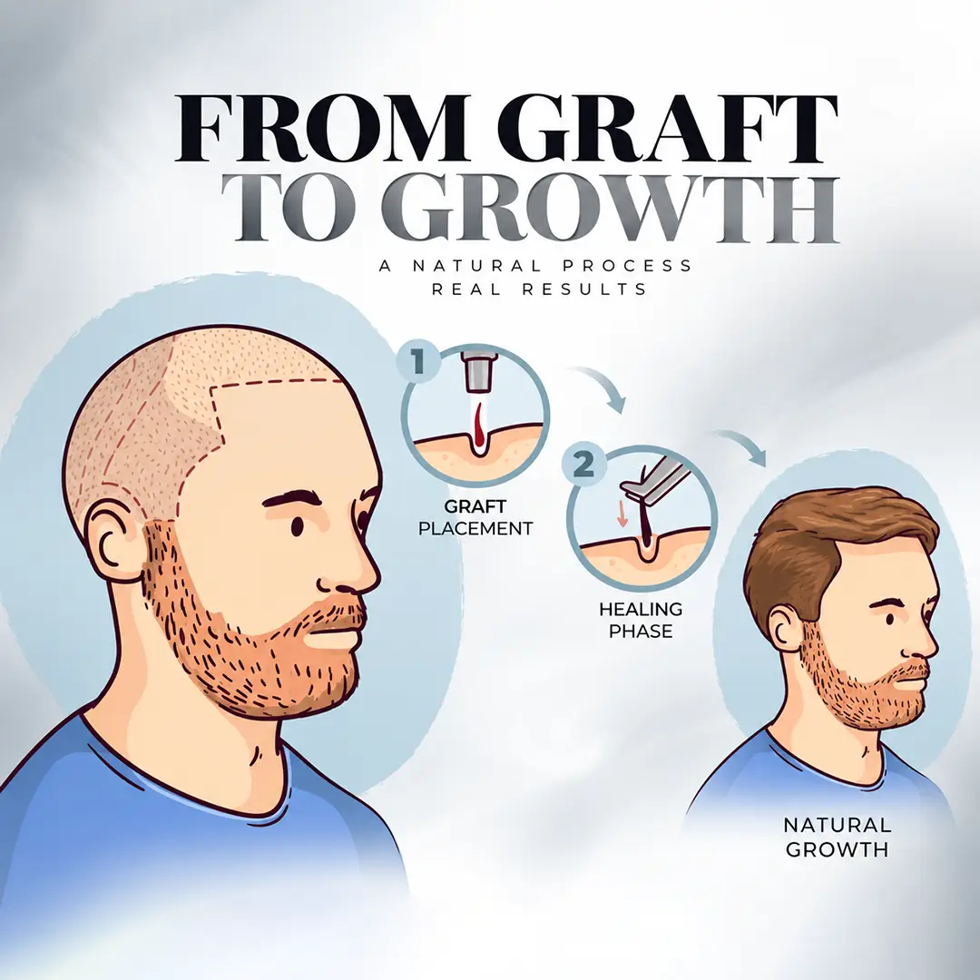
The recovery timeline is a predictable sequence of five stages: immediate healing in week 1; shedding across weeks 2 to 6; a dormant stretch through months 2 and 3; visible growth from month 4; maturation continuing for 18 months. Sequence rarely changes even though speed does.
Week 1 brings crusting plus mild swelling around the recipient zone. Weeks 2 to 6 bring the shed. Months 2 and 3 look flat, which is why the stretch earned the nickname ugly duckling phase. Month 4 delivers the first visible sprouts. Months 6 to 9 add calibre as thin vellus shafts thicken into terminal hair. For example, a 3,500-graft frontal case usually reads convincingly at month 8 while the same graft count spread across a crown reads considerably later. Our coordinators schedule photo reviews at month 3, month 6, month 12. Smaller zones such as an eyebrow transplant mature faster because the hairs are shorter.
| Stage | Window | What the photograph shows |
|—|—|—|
| Healing | Days 1 to 10 | Crusting, mild swelling, transplanted shafts visible |
| Shedding | Weeks 2 to 6 | Transplanted shafts fall out, scalp looks emptier |
| Dormant | Months 2 to 3 | No visible change, the ugly duckling phase |
| Early growth | Months 4 to 6 | Fine sprouts appear, density still thin |
| Thickening | Months 6 to 9 | Shafts gain calibre, hairline reads clearly |
| Maturation | Months 9 to 18 | Final density, crown completes last |
Windows above describe typical progression, not a promised schedule.
Why Does Transplanted Hair Shed After Surgery?
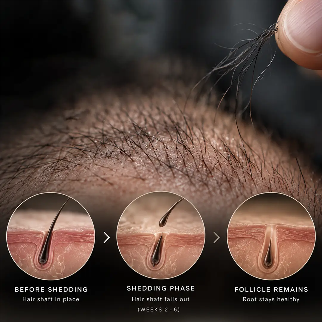
Post-transplant shedding is a normal telogen response in which the transplanted shaft falls out while the follicle stays anchored in the scalp. The shed begins around week 2 and runs to week 6. Losing up to 90% of visible shafts during the phase does not signal failure.
Biology explains the pattern. Surgical handling pushes relocated follicles into a resting phase so the shaft detaches while the root survives. Loh and colleagues documented localised telogen effluvium after hair transplantation in Annals of Dermatology (2018). Guerrero-González and colleagues mapped the same effect in the donor area using Trichoscopy in 2023. Shock loss affecting surrounding native hair is a separate event, usually temporary. For instance, a patient with dense native hair around the recipient zone sheds more visibly than a fully bald recipient area. Our nursing team warns every patient before discharge so month 2 photographs cause no alarm. A beard transplant follows the same shedding curve.
Why Does Crown Before and After Lag Behind the Hairline?
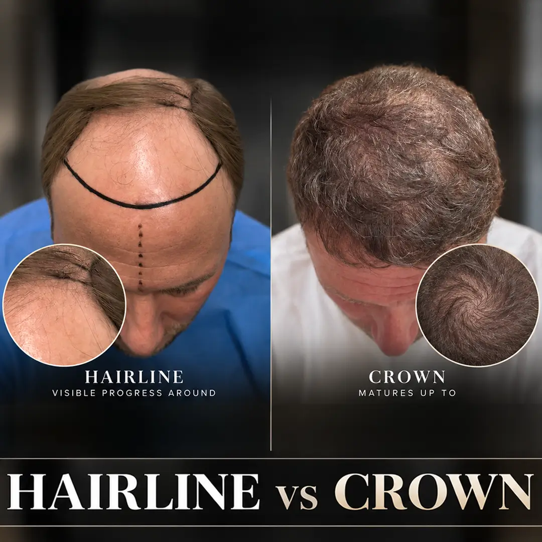
The crown is the slowest zone in any hair transplant before and after comparison because whorl geometry scatters light and shallow exit angles reduce apparent density. Hairline results usually read at month 12 while crown results keep improving for 18 months. Crown coverage also competes with ongoing native miniaturisation.
Geometry drives the difference. Frontal hairlines sit on a flat plane where each graft covers visible surface efficiently. The crown radiates outward from a central point. Identical graft numbers spread across a curved area there and reflect light unevenly. According to Queen and colleagues in Dermatologic Surgery (2025), zone-specific planning separates realistic outcomes from disappointing ones. Crown work suits a stable donor supply but rarely suits a patient with Norwood stage 3 loss because pattern loss keeps expanding and leaves an island of transplanted hair. In practice, our surgeons treat the frontal third first, then reassess the crown at month 12. Technique choice matters here too as the guide to DHI implantation explains.
How Should Before and After Photos Be Documented?
A reliable before and after record is a fixed protocol rather than a casual snapshot: same camera; same distance; same lighting; same five angles; dry unstyled hair. Photographs captured at day 0, month 3, month 6, month 12 build a comparable series across 12 months.
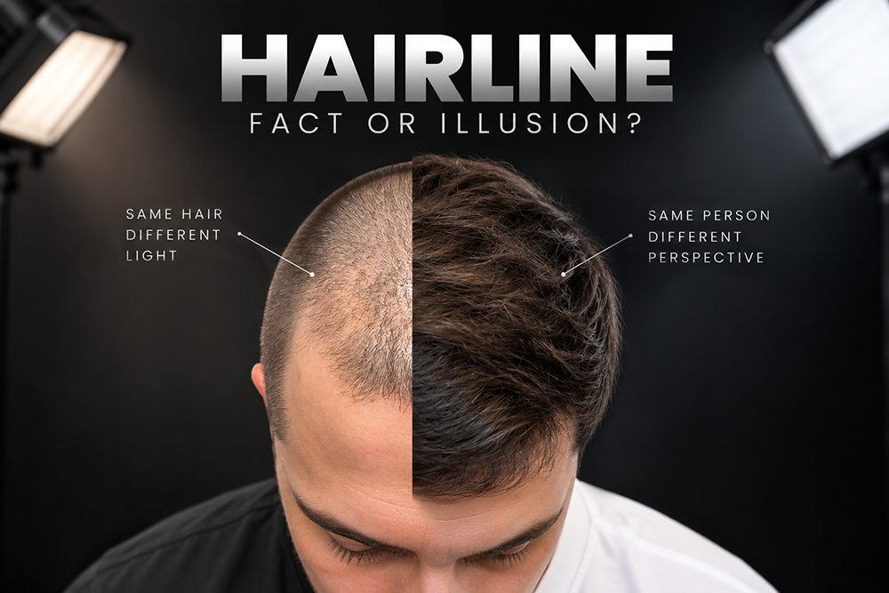
Protocol beats equipment. First, a phone camera used consistently outperforms a professional shoot repeated once. Five angles matter: front; left profile; right profile; mid-scalp from above; crown from behind. Hair should be dry and unstyled. Product or wet shafts both exaggerate density badly. For example, a crown photographed under a ceiling downlight looks thinner than the same crown under diffuse daylight. Our photography protocol fixes camera height alongside a neutral background at every visit, which is what makes month 3 comparable to month 12. Patients travelling home replicate the setup with International Plus guidance sheets, a routine described in our decision framework for medical travel.
How Do You Spot an Unreliable Before and After Photo?
An unreliable before and after photo is a frame where something other than hair growth changed between the 2 images. Lighting shifts; hairstyle changes; wet versus dry hair; camera angle; crop: each inflates apparent density. Missing donor area shots hide over-harvesting completely.
Reading a gallery critically takes four checks. First, look for the donor area in at least one image since over-harvesting shows at the back rather than the front. Second, confirm the time stamp because a result photographed at month 6 will improve further. Third, check whether the hairstyle changed between frames. Fourth, look for multiple time points rather than a single dramatic pair. Liu and colleagues linked transplantation satisfaction to self-esteem outcomes in 2019, which is why misleading galleries carry genuine consequence. Real-world documentation includes the awkward months. Our clinic keeps month 3 images even when the frame flatters nobody. Verification habits appear in our overview of the most trusted clinics in Turkey.
When Should Slow Growth Be Investigated?
Slow growth is a clinical question when a transplanted zone shows no visible sprouting by month 6. Density stalling between month 9 and month 12 also warrants review. Redness, pustules or persistent numbness at any point deserve prompt examination. Most cases resolve as normal timeline variation rather than as graft failure.
Assessment beats waiting. A physician examines the recipient zone under magnification, checks the donor area, then reviews the standardised photo series against expected timelines. Kwack and colleagues in Dermatologic Surgery (2021) studied the factors governing graft survival, which is why handling questions belong in any late review. Medical support for native hair is assessed by the examining physician, not chosen online. In practice, our follow-up team compares month 9 images against the day 0 baseline before discussing a second procedure. Reading hair transplant before and after results properly means judging the plan against the calendar. Women should check our guide to female candidacy; budgets sit in the hair transplant cost breakdown. International Plus holds a 4.8/5 rating over 5,504 verified reviews. Individual results vary.
Book a photo based progress review with our coordination team and receive a standardised documentation schedule for your own case.
Frequently Asked Questions
When will I see final hair transplant before and after results?
Final results usually read between month 12 and month 18. The hairline typically matures around month 12 while the crown continues thickening for another 6 months. Photographs taken before month 9 understate the outcome because shafts are still gaining calibre.
Is it normal to lose transplanted hair after surgery?
Yes. Transplanted shafts shed between weeks 2 and 6 in nearly every case, sometimes losing 90% of what was visible on the operating day. The follicle stays anchored, and regrowth begins around month 4.
Why does the crown take longer than the hairline?
Crown hair grows from a whorl that radiates outward so the same graft count spreads across a curved surface and reflects light unevenly. Shallow exit angles reduce apparent density further. Hairline zones sit on a flat plane, so frontal results read at month 12 while crown results need month 18.
What should results look like at month 3?
Month 3 usually looks flat or slightly thinner than the pre-surgical baseline, which is why clinicians call it the ugly duckling phase. Transplanted shafts have shed, native hair may have thinned temporarily, new growth has not surfaced. Month 3 images are documentation, not an outcome preview.
Can before and after photos be trusted?
Only when the series is standardised. Look for consistent lighting, an unchanged hairstyle, dry hair, multiple time points, plus a donor area frame. A single dramatic pair under different lighting proves little. Reputable clinics publish awkward interim months too.
Do women follow the same recovery timeline?
Broadly yes though visible improvement often reads later in women because diffuse thinning surrounds the transplanted zone rather than framing a defined bald patch. Native hair around the grafts can also shed temporarily. Documentation across month 6, month 12, month 18 matters more for female patients.
Reviewed by Dr. Eren Gülbahar
References
- Fu D, et al. Plast Reconstr Surg. 2026. PMID: 40690367
- Loh SH, et al. Ann Dermatol. 2018. PMC5839894
- Guerrero-González GA, et al. Skin Appendage Disord. 2023. PMC10601894
- Queen D, et al. Dermatol Surg. 2025. PMID: 40354670
- Liu F, et al. J Cosmet Dermatol. 2019. PMID: 30582267
- Kwack MH, et al. Dermatol Surg. 2021. PMID: 33165067
Follow us on Social Media
internationalplusofficial
internationalplus
internationalplusdental
This content is written, reviewed, and approved by the International Plus Medical Review Board to ensure clinical accuracy and adherence to strict editorial standards. All medical information is regularly monitored, audited and updated in light of the latest scientific advancements.
However the information provided here is for general informational purposes only and cannot be used for self diagnosis or making individual health interpretations. Results of medical treatments depend on individual anatomy and the unique healing process of each patient. This information should not replace a personal consultation with a qualified healthcare professional. To understand the best options for your specific needs and to receive a personalized treatment plan, we invite you to book a free consultation with the expert medical team at International Plus.

I am a healthcare professional with over 4 years of experience in dentistry, specializing in prosthetic dental treatment and implant-based applications. Since 2024, I have been serving as chief physician, leading clinical management, team coordination, and international patient processes. With my B2-level English proficiency, I am able to communicate effectively with international patients. I am a patient-centered, innovative, and solution-oriented dentist who prioritizes patient satisfaction.