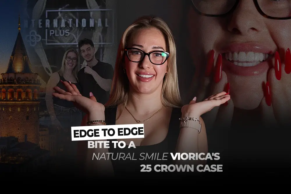

An edge to edge bite is a bite in which the upper and lower front teeth meet tip to tip instead of the upper teeth overlapping the lower ones. Viorica, a 28 year old patient from Romania, came to International Plus because her front teeth met this way and also overlapped one another. Her record lists a Class II bite with the front teeth meeting edge to edge. She told the team on the first day that her aesthetic expectations were very high. This case follows how her treatment was planned and carried out from two infected molars and thin sinus bone to 25 crowns finished with natural translucent edges.
Key Takeaways
- Viorica’s complaint was aesthetic: an edge to edge bite plus overlapping front teeth, recorded as a Class II bite.
- Two infected upper molars were removed, an open sinus lift was carried out on one side because the bone measured less than 4 mm and 2 implants were placed.
- She was never without teeth: composite temporaries were made on day one and PMMA temporaries were fitted on day two.
- Her back teeth were kept so the vertical dimension of the bite was not changed.
- An implant plan needs 4 to 6 months of healing before the final implant crowns but Viorica chose it because the record notes she wanted the longer lasting option.
Viorica’s Story: High Expectations and a Bite She Wanted Changed
Viorica’s main concern was the look of her smile, not pain. Her record describes a young woman with high aesthetic expectations who was unhappy with an edge to edge bite and overlapping front teeth. The problem was not serious enough to affect her social life. Like many young adults with a strong aesthetic sense, Viorica had a clear picture of the smile she wanted.
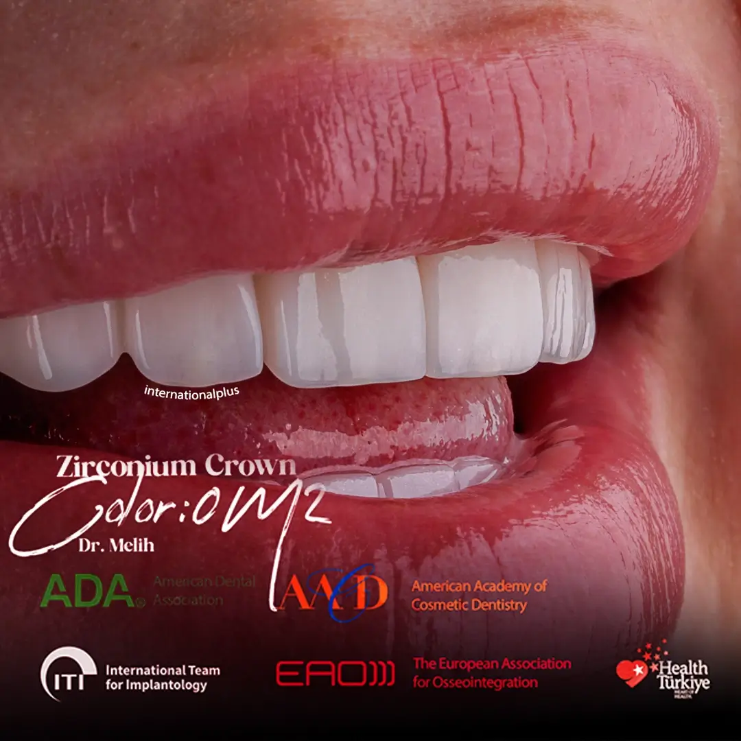
She arrived slightly anxious according to the team’s notes but hopeful about the result. Her request was specific. She wanted a smile that fills the mouth with translucent incisal edges and a completely natural look rather than a flat white finish. The design stayed with that brief from the first scan to the final try in. For a patient with expectations this precise, the planning has to show the result before any tooth is prepared, which is why her case was planned digitally from the start with digital smile design preview technology. For example, the translucency she asked for is decided at the design stage, not added at the end.
What the Examination Found
The examination was the first step in a digital workflow. Panoramic images and radiographs showed more than the aesthetic problem she came with: infected molars in the upper jaw, sinus bone less than 4 mm thick at the site and uneven gum levels in the lower jaw.
Several plans were offered to her. Some used implants, others did not. Viorica chose the implant route because it was the longer lasting option. In Viorica’s case the findings fell into three groups: first the infected upper molars, second the thin sinus bone, third the uneven gum levels. A Trios 5 intraoral scan was part of her record. According to a 2023 network meta analysis by Vitai et al., complete arch accuracy differs between intraoral scanners and between clinical situations so the scanner chosen for a full arch case matters. The findings are summarised below.
| Finding | What was done |
| Edge to edge bite, overlapping front teeth | Crown design planned on a full digital and face scan record |
| Infected upper molars | 2 upper teeth extracted and the infection cleaned |
| Sinus bone under 4 mm | Open sinus lift on one side, then 2 implants |
| Uneven upper gum line | 3 gingivectomies in the front of the upper jaw |
| Uneven lower gum levels | Pink porcelain to even the visible gum line |
| Additional treatment in the record | 6 root canal treatments, decay removal and fillings |
Bottom line: the aesthetic request was planned alongside separate treatment for the infection and the thin bone, plus the gum levels.
The Surgical Phase: Extractions, Open Sinus Lift and Two Implants
An open sinus lift, also called a lateral window sinus lift, raises the floor of the sinus through a small window in the side of the upper jaw so new bone can form under the membrane. The team chose the open approach because the remaining bone was thinner than 4 mm, which is too little for a simpler closed lift.
Two infected upper teeth were extracted first. The infection was cleaned before the sinus was lifted on one side. Then 2 implants were positioned in the upper jaw. According to a 2019 systematic review by Raghoebar et al., lateral window sinus augmentation in jaws with residual bone of 6 mm or less showed an annual implant loss of 0.43% over at least five years. The record plans 4 to 6 months for osseointegration, the process where bone fuses to the implant surface, which Viorica will spend at home in Romania. Patients reading about the wider procedure can compare Martina’s case of sinus lift dental implants for a full mouth reconstruction and our page on dental implant treatment in Istanbul.

Temporaries, Gum Contouring and the Digital Design
Viorica left the clinic with teeth on the first day. A silicone impression of her own natural teeth was used to make composite temporaries in the chair. On the second day she received PMMA temporaries, made from the same impression in a stronger acrylic and tried in for comfort and appearance.
A 2022 systematic review by Jain et al. concluded that 3D printed provisional resins can serve as an alternative to milled or conventional long term temporaries. During the temporary phase, 3 gingivectomies, small reshaping procedures that remove excess gum, levelled the front of the upper gum line. She returned the next day for a check and the bite was adjusted so the temporaries met evenly. The final design was built from a Trios 5 intraoral scan plus a face scan so the new teeth would sit symmetrically in relation to her face. A 2025 single participant study by Al Hamad et al. found a face scan workflow feasible for treatment planning and smile design but not reliable for manufacturing demanding prostheses. Photogrammetry, a camera system for recording implant positions, was not needed because no full arch implant framework was planned. In practice, Viorica tried her new smile in temporary form before the final crowns were made.
The Final Restoration: 25 Crowns, Pink Porcelain and a Natural Finish
The final restoration was 25 crowns in shade 0M2. Our in house laboratory team designed and layered the crowns, which were cemented with GC FujiCem. The treatment plan also included six root canal treatments along with decay removal plus fillings. Because her back teeth were kept, the vertical dimension of her bite did not change.
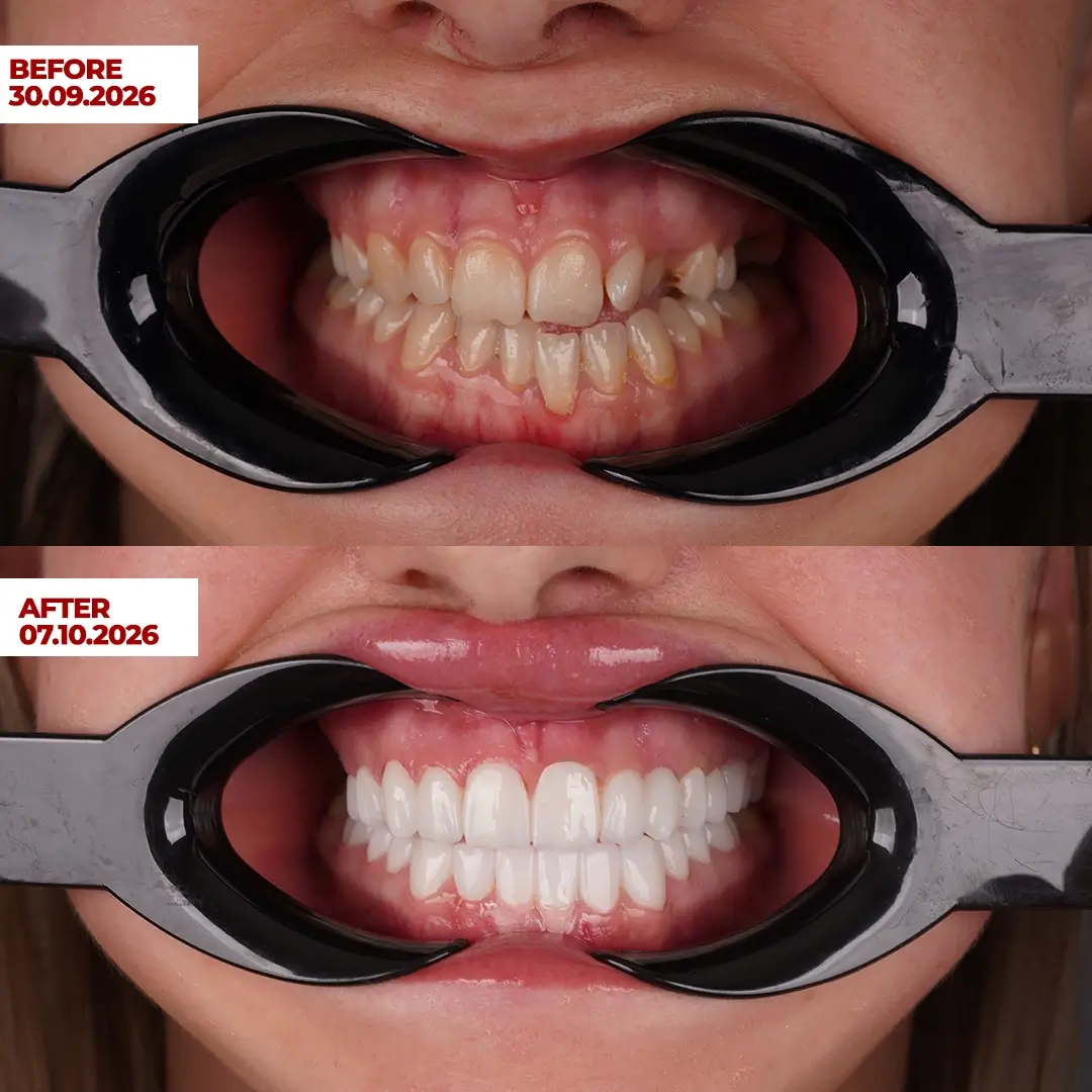
In the lower jaw the gum levels were uneven so pink porcelain was used to make the teeth appear to rise from one level line. A 2018 technique report by Papaspyridakos et al. describes pink ceramics in anterior implant bridges as a way to reduce the need for extensive soft and hard tissue surgery in the upper aesthetic zone. Three try ins took place between 1 and 7 September. The record notes that the edge to edge bite was corrected. When she returns after healing, one digital scan will record both implants and the final 2 implant crowns will bring the total to 27 units. Readers can see another case combining root canal treatment with crowns in Ornella’s root canal before crown case and the crown options on our dental crowns treatment page.
The Result and Viorica’s Experience
Viorica left very satisfied. Her record says she described the clinic as professional and the process as one she was pleased with. The smile matches the brief she gave on the first day: a full, natural design with translucent edges.
The case began with a digital scan and finished with a digital scan and the same records will guide her implant crowns. A 2017 paper by Kuljic describes edge to edge incisor positions as an aesthetic challenge where a careful risk assessment guides the treatment plan. International Plus has treated more than 200,000 patients from 140+ countries since 2014 and holds 5,504 verified reviews at 4.8 out of 5. This case was reviewed for accuracy by our clinical team. Individual results may vary. This case study is educational and is not medical advice.
If your front teeth meet edge to edge and you want a natural looking result, Book Your Free Consultation and send photographs of your smile to our coordinators. To discuss implants and an open sinus lift in the same trip, Contact our patient coordination team for a no obligation review.
Frequently Asked Questions
What is an edge to edge bite?
An edge to edge bite is a bite in which the upper and lower front teeth meet tip to tip when you close, instead of the upper teeth sitting slightly in front of the lower ones. It can make front teeth look short or worn and concentrates biting force on the edges. A dentist assesses it with photographs and scans, plus a bite record.
Can crowns correct an edge to edge bite?
In some adults the appearance of an edge to edge bite can be changed with crowns when the plan is made on a full digital record and the back teeth support the bite. Other cases need orthodontics or surgery. The right route depends on the jaw relationship and the condition of the teeth. The patient’s goals matter too so the route is decided after examination.
What is an open sinus lift?
An open sinus lift, also called a lateral window sinus lift, raises the sinus floor through a small window in the side of the upper jaw so bone can form for an implant. It is usually chosen when the remaining bone under the sinus is thin and implants then heal for several months before crowns are fitted.
Will I have teeth while my implants heal?
In most planned cases patients leave with temporary teeth. In this case composite temporaries were made on the first day from an impression of the patient’s own teeth and stronger PMMA temporaries followed on the second day. The final implant crowns are fitted after the healing period, usually several months later.
What is pink porcelain used for?
Pink porcelain is a gum coloured ceramic added to crowns or bridges to make uneven gum levels look even. It can hide differences in gum height without further gum surgery in suitable cases. It is shaded to match the patient’s own gums so the crowns appear to emerge from one natural, level gum line.
Reviewed by Dr. Eren Gülbahar
References
- Raghoebar GM, et al. J Clin Periodontol. 2019. PMID: 30624789
- Jain S, et al. Polymers (Basel). 2022. PMID: 35808735
- Al Hamad KQ, et al. J Prosthodont. 2025. PMID: 39837798
- Papaspyridakos P, et al. J Prosthodont. 2018. PMID: 27037907
- Kuljic B. Compend Contin Educ Dent. 2017. PMID: 28257224
- Vitai V, et al. J Dent. 2023. PMID: 37516338
Follow us on Social Media
internationalplusofficial
internationalplus
internationalplusdental
This content is written, reviewed, and approved by the International Plus Medical Review Board to ensure clinical accuracy and adherence to strict editorial standards. All medical information is regularly monitored, audited and updated in light of the latest scientific advancements.
However the information provided here is for general informational purposes only and cannot be used for self diagnosis or making individual health interpretations. Results of medical treatments depend on individual anatomy and the unique healing process of each patient. This information should not replace a personal consultation with a qualified healthcare professional. To understand the best options for your specific needs and to receive a personalized treatment plan, we invite you to book a free consultation with the expert medical team at International Plus.

I am a healthcare professional with over 4 years of experience in dentistry, specializing in prosthetic dental treatment and implant-based applications. Since 2024, I have been serving as chief physician, leading clinical management, team coordination, and international patient processes. With my B2-level English proficiency, I am able to communicate effectively with international patients. I am a patient-centered, innovative, and solution-oriented dentist who prioritizes patient satisfaction.