3D smile design is a planning method that merges a facial scan with an intraoral scan plus a cone beam CT into one aligned three dimensional model, then converts that model into physical objects a patient can wear and chew on. At International Plus the facial capture takes 10 seconds at 50 micron resolution and the aligned file then drives a printed mock up, a preparation guide, a milled restoration. This guide does not describe what 3D smile design looks like on a screen. This guide follows the design out of the software and into the mouth, stage by stage, including the points where accuracy is lost.
Key Takeaways
- A virtual patient is the aligned combination of three datasets: face, teeth plus jawbone. No single scanner produces the virtual patient alone.
- The printed mock up and silicone index are the two objects that carry screen geometry into the mouth; without them a 3D design stays a picture.
- A five axis milling unit finishes zirconia to micron level fit and sintering at 1500 to 1800 degrees Celsius shrinks the framework by a calculated factor the software compensates for in advance.
- Digital workflows gain speed and repeatability but a capture error at the scanning stage travels down the entire production chain into the final restoration.
- The design file is archived so a fractured unit can be remanufactured years later against the approved geometry rather than redesigned from scratch.
What Is 3D Smile Design and Which Data Does the Design Need?
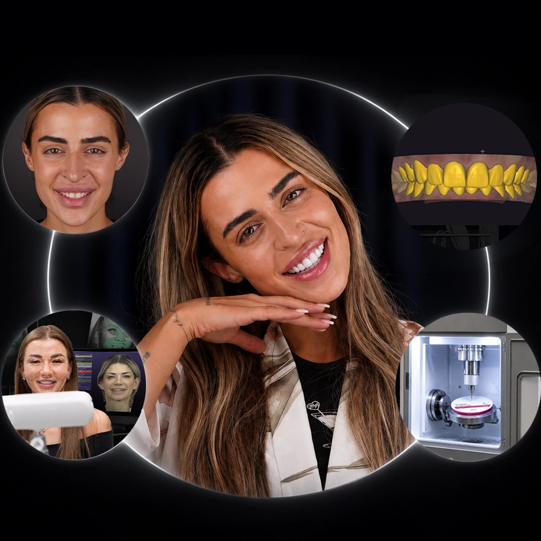
3D smile design needs three separate captures before any tooth shape is drawn. A SHINING 3D MetiSmile facial scanner records the face in 10 seconds at 50 micron resolution, mapping lip line and facial midline plus the way the cheeks frame the smile. A TRIOS 5 intraoral scanner on the 3Shape platform records teeth and gingiva in the same session. A cone beam CT supplies root position and bone volume, the layer no surface scanner reaches. Alignment of the three datasets produces what the literature calls the virtual patient and a 2022 review in BMC Oral Health described prosthetic set up on such merged datasets as the basis of modern virtual planning (Flügge et al., 2022). Our Diagnostic Council reviews the merged file before design work starts because a misaligned CBCT overlay produces a beautiful design over a root that cannot support the restoration. Alignment is verified against three fixed anatomical landmarks rather than a single reference point.
Why Does a 3D Design Differ From a Flat 2D Smile Plan?
A 2D plan draws outlines on a photograph. A 3D design carries depth, angle and volume, which changes four decisions the 2D plan cannot make. Every dental smile design choice that involves depth needs that third axis. Incisal edge position becomes a measured coordinate rather than an estimated line. The occlusal plane can be checked against the interpupillary line and the horizon of the face at the same time. Facial midline is read from the aligned facial scan instead of the nose, which frequently sits off centre. Speech and smile dynamics enter the plan because the scan records the face in repose and in a wide smile so tooth length is tested against lip travel before ceramic exists. Patients comparing preview formats find the practical differences set out in our guide to smile simulation accuracy. The design leaving our software is a coordinate set rather than an illustration and the production chain reads coordinates.
Learn more about our DSD planned smile transformation protocol before booking a scan appointment.
How Does the Design Travel From the Screen Into the Mouth?
Transfer happens through physical intermediates, each manufactured from the same approved file. The printed model reproduces the proposed arch in resin. The silicone index, moulded over that model, carries the design outline back onto the untouched teeth as a resin trial smile. The preparation guide, printed from the difference between existing teeth and the planned outline, tells the clinician exactly how much enamel the design requires and where the design requires none. Only then does milling begin. A 2024 meta analysis in the Journal of Prosthetic Dentistry compared 3D printed against milled and conventionally processed prosthetic materials and reported material behaviour differences that justify printing provisionals while milling definitive units (Valenti et al., 2024). In our practice the trial smile is worn for at least 48 hours before preparation because a design that survives two days of eating and speaking has been tested rather than admired.
| Stage | What the stage produces | Accuracy or time | What the patient does |
|—|—|—|—|
| 3D data capture | Facial scan, intraoral scan, CBCT dataset | Facial capture in 10 seconds at 50 microns | Sits for three short scans, no impression trays |
| Virtual patient alignment | One merged file holding face, teeth plus jawbone | Same day, verified on three landmarks | Nothing; the Diagnostic Council reviews the merge |
| 3D design | Tooth coordinates, occlusal plane, incisal edge | Design session plus revision round | Reviews the proposal and requests changes |
| Printed mock up and index | Resin model, silicone index, trial smile | Printed overnight, worn 48 hours or longer | Wears and tests the trial smile, then approves |
| Preparation guide | Printed reduction guide from the approved design | Chairside use during preparation | Attends the preparation appointment |
| Milled restoration | Zirconia or ceramic units from the in house lab | Five axis milling, sintering 1500 to 1800 C | Attends try in and fitting appointments |
Bottom line: every row after the first depends on the accuracy of the row above, which is why capture quality dictates the quality of the finished teeth.
What Does 3D Printing Actually Produce in Smile Design?
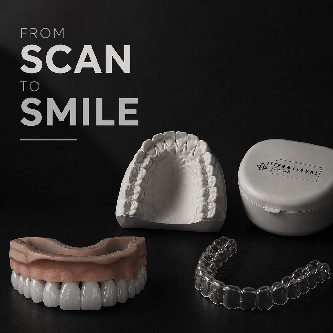
3D printing produces the rehearsal, not the result. An Asiga printer with Arma dental resin builds the study model, the trial smile shell plus the preparation guide from the approved design file. Printed PMMA provisionals then carry the design through the healing and adaptation period while the gum settles around new contours. Printed objects are not the definitive teeth and the distinction is a material one: a 2024 systematic review in the Journal of Dentistry comparing printed and conventionally milled zirconia reported differences in trueness, precision as well as surface behaviour between the two production routes (Alghauli et al., 2024). Printing is fast and iterative, cheap enough to repeat, which is precisely what a rehearsal stage should be. Our technicians print a fresh model for every design revision rather than adjusting an old one since a modified model no longer matches the digital file it is supposed to represent.
How Is the Final Restoration Milled and Finished In House?
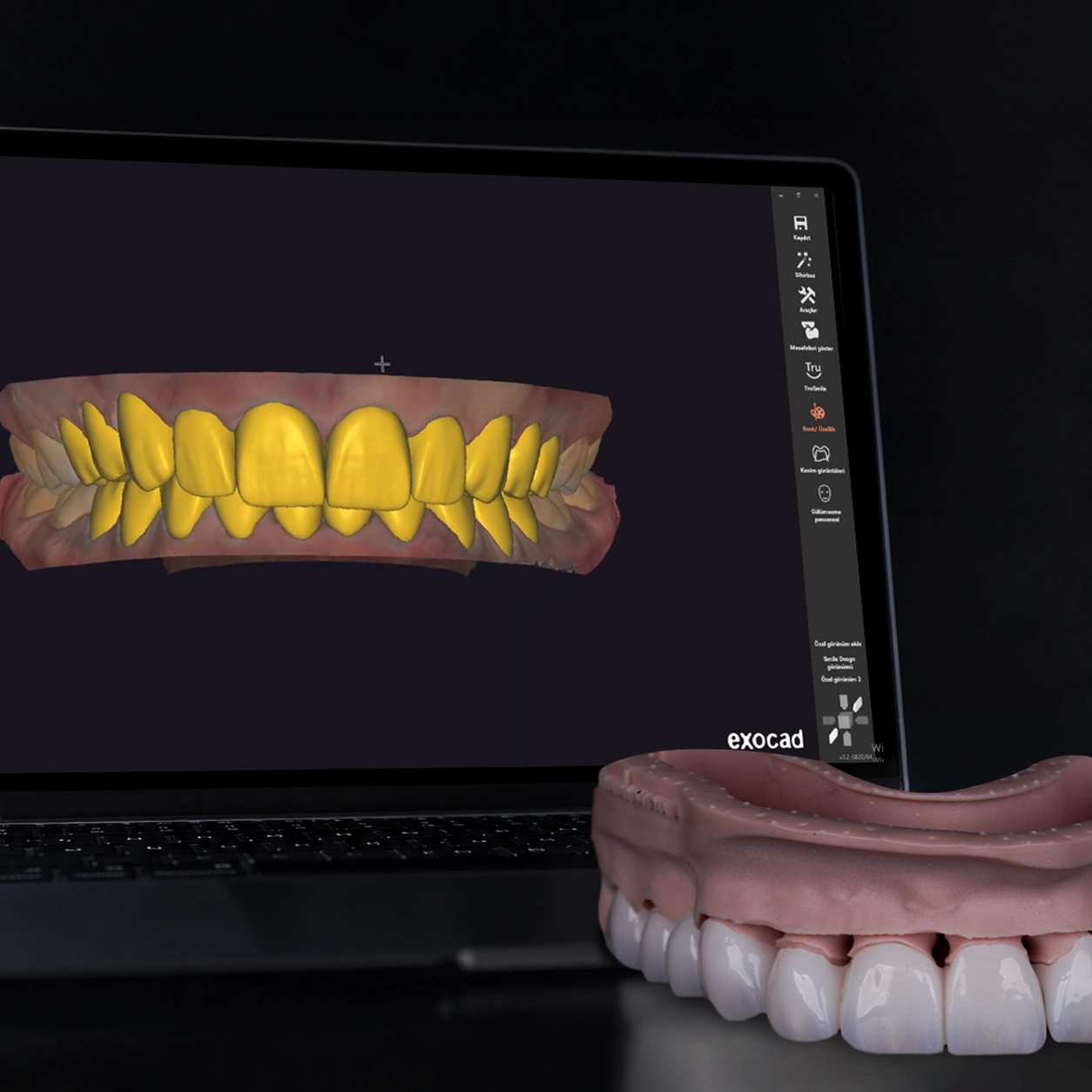
The definitive units are subtractive, cut from a solid disc rather than built up in layers. A Redon five axis milling unit reads the approved design file and cuts the framework to micron level tolerances. Sintering follows at 1500 to 1800 degrees Celsius, during which zirconia shrinks by a known percentage the milling software has already compensated for in the enlarged pre sintered geometry. Final glaze firing runs at 800 to 900 degrees. Ivoclar zirconia is our standard for definitive units and multilayer discs supply the value gradient from cervical to incisal. Because the laboratory sits inside the clinic, a try in adjustment noted at 10:00 can be milled and returned the same afternoon, a turnaround that matters when a patient has flown in for a fixed number of days. A 2026 systematic review and meta analysis in the Journal of Prosthodontics assessed the cost efficiency of digital against conventional workflows and found the digital route reduced clinical and laboratory time (Muehlemann et al., 2026). The same file that produced full ceramic crown restorations also documents them.
Book Your Free Consultation → Talk to our smile design coordinators on WhatsApp
Why Does the Archived Design File Matter Years Later?
An approved design file is a manufacturing record with a long service life. When a single unit fractures after five years, the archived geometry allows the laboratory to mill a replacement against the original approved coordinates instead of reverse engineering a shape from a worn adjacent tooth. Shade, contour, contact points plus emergence profile are all recorded in that file. Patients planning a digital smile makeover who ask how long veneers last are usually asking a second question underneath: what happens when one unit fails. Archiving answers it. The file also documents what was approved, which protects the patient in any later discussion about whether the delivered restoration matched the agreed design. Our full mouth cases, including the couple treated with full mouth zirconia crowns, keep the approved design alongside the delivered outcome in the same patient record. Individual results may vary and stored geometry is not a warranty of tissue behaviour.
Where Does the 3D Smile Design Workflow Reach Its Limits?
A digital chain gains speed and repeatability but a capture error committed at the scanning stage travels untouched into the printed guide and the milled restoration. A 2024 systematic review in the International Journal of Implant Dentistry examined the factors that degrade intraoral scanning accuracy, including arch length, scan strategy as well as reflective surfaces (Gehrke et al., 2024). Facial scanners carry their own tolerance: a 2020 study in the Journal of Prosthetic Dentistry measured trueness and precision of a dual structured light facial scanner and reported measurable interexaminer variation (Piedra-Cascón et al., 2020). Soft tissue is the second limit since gum margins move for weeks after contouring and no scan predicts that migration exactly. Patient compliance is the third: a trial smile removed after two hours has not been tested. In our practice we re scan the arch whenever the printed model rocks under finger pressure because a model that does not seat stably indicates a distorted scan rather than a printing fault. Reading a phone based preview against these tolerances is covered in our review of what a smile design app can and cannot capture.
Getting 3D Smile Design Right From Scan to Fitting
Judging a 3d smile design proposal means asking about production, not only about the picture. Ask which three datasets were captured. Ask whether a physical mock up will be produced, who signs off the merged file plus where the milling happens. Dr. Eren Gülbahar, Head of Cosmetic Dentistry and an American Dental Association member, calls the trial stage a test drive of the smile and roughly 3,000 international patients have gone through that stage under his care. International Plus combines official Turkish Ministry of Health authorization, 4.8 out of 5 across 5,504 verified reviews, more than 200,000 patients treated from 140+ countries since 2014 and an in house laboratory that mills what our software designed, which makes the chain from scan to fitting auditable at every step. Our broader account of golden ratio smile planning covers the aesthetic reasoning behind the coordinates. Individual results may vary and a personalised clinical examination remains the only way to establish what a specific mouth can support.
Discover 3D Smile Design at International Plus → See how a full smile transformation is planned
Frequently Asked Questions
How long does a 3D smile design take from scan to finished teeth?
Scanning occupies a single appointment with facial capture taking 10 seconds. Design and Diagnostic Council review follow within days. Printed mock up approval, preparation plus milling of definitive units are usually completed inside one treatment trip for international patients. Complex cases requiring gum healing need a second visit weeks later.
Is 3D smile design more accurate than a 2D digital design?
A 3D design records depth, occlusal plane plus root position so tooth length and angle become measured values rather than estimates on a photograph. Accuracy still depends on capture quality since scanning error carries forward into production. Neither format removes the need for a physical trial smile before preparation begins.
Does a digital 3D design require tooth preparation?
Preparation depends on the design, not on the technology. A printed reduction guide made from the approved file shows exactly where enamel must be removed and where existing tooth shape already matches the plan. Additive cases with adequate space sometimes need no reduction at all, which the guide makes visible before treatment.
What is a virtual patient in 3D smile design?
A virtual patient is one aligned digital model combining a facial scan with an intraoral scan plus a cone beam CT. Face supplies proportion and lip dynamics. The intraoral scan supplies tooth and gum surfaces while the CBCT supplies root and bone position. Design decisions made on the merged model account for all three layers.
Can the same 3D design be used again if a tooth breaks?
Yes provided the design file was archived. The laboratory mills a replacement unit against the original approved coordinates, matching contour, contact points plus emergence profile without redesigning the restoration. Shade may need rechecking against neighbouring units that have aged. Individual results may vary and a clinical examination determines whether replacement alone is appropriate.
Reviewed by Dr. Eren Gülbahar
References
- Flügge T, et al. BMC Oral Health. 2022. PMC8802526
- Valenti C, et al. J Prosthet Dent. 2024. PMID: 35934576
- Alghauli M, et al. J Dent. 2024. PMID: 38471580
- Muehlemann E, et al. J Prosthodont. 2026. PMC12990028
- Gehrke P, et al. Int J Implant Dent. 2024. PMC11063012
- Piedra-Cascón W, et al. J Prosthet Dent. 2020. PMID: 31918895
Follow us on Social Media
internationalplusofficial
internationalplus
internationalplusdental
This content is written, reviewed, and approved by the International Plus Medical Review Board to ensure clinical accuracy and adherence to strict editorial standards. All medical information is regularly monitored, audited and updated in light of the latest scientific advancements.
However the information provided here is for general informational purposes only and cannot be used for self diagnosis or making individual health interpretations. Results of medical treatments depend on individual anatomy and the unique healing process of each patient. This information should not replace a personal consultation with a qualified healthcare professional. To understand the best options for your specific needs and to receive a personalized treatment plan, we invite you to book a free consultation with the expert medical team at International Plus.

I am a healthcare professional with over 4 years of experience in dentistry, specializing in prosthetic dental treatment and implant-based applications. Since 2024, I have been serving as chief physician, leading clinical management, team coordination, and international patient processes. With my B2-level English proficiency, I am able to communicate effectively with international patients. I am a patient-centered, innovative, and solution-oriented dentist who prioritizes patient satisfaction.