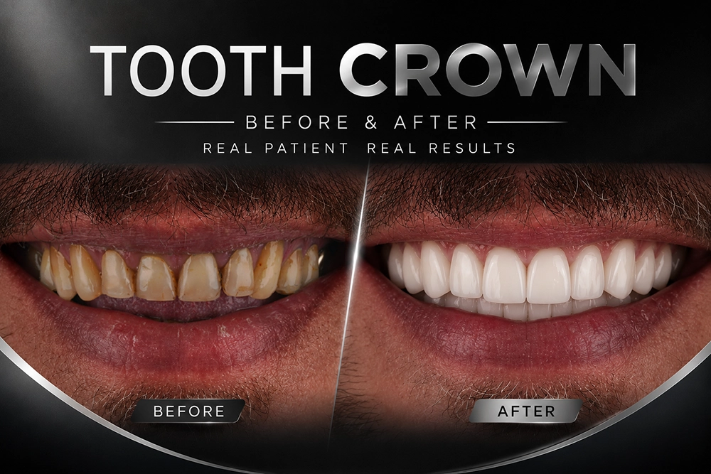
Around 12,000 searches a month in the United States go to crown result images, and almost every one of those searchers wants the same answer: is the change real, does the tooth still read as a tooth. Tooth crown before and after outcomes vary far more than a polished gallery suggests because the visible difference depends on which tooth is treated and how dark the starting shade was. Gum health sets the ceiling on the rest. This guide explains what a crown changes, what a crown leaves untouched, then how to read a result photograph without being misled. Individual results may vary and a personalized consultation remains the only way to assess a specific tooth.
Key Takeaways
- A crown changes colour, contour and surface texture; a crown does not move teeth, whiten neighbours or restore a receded gum margin.
- The most dramatic tooth crown before and after difference appears on a single darkened upper front tooth, where a shade shift of four or more VITA steps is common.
- Shade matching is measured, not guessed: our workflow uses a TRIOS 5 intraoral scan plus a MetiSmile face scan at 50 micron resolution before any design begins.
- A 2018 systematic review by Pjetursson and colleagues recorded 5 year survival above 95% for single ceramic and metal ceramic crowns so the aesthetic question usually outweighs the durability question.
- Comparable photographs need the same angle, retraction and lighting; heavy editing can add two apparent shade steps that daylight will not show.
What Actually Changes Between a Tooth Crown Before and After?
A dental crown is a full coverage restoration that replaces the entire visible surface of a damaged tooth, so the recorded difference is a change in colour and contour, plus a change in surface texture. Tooth position does not change. Tooth alignment and gum level stay as they were, and lip support stays unchanged unless separate treatment is planned alongside the crown. A 2018 systematic review of single crowns by Pjetursson and colleagues reported 5 year survival above 95% for zirconia ceramic and metal ceramic units, which is why aesthetics, not survival, dominates most crown consultations at International Plus. In our clinic the visible gains patients notice first are a shade shift of two to four VITA steps and closure of a chipped incisal edge, with tooth width restored inside the smile line. The crown will not lighten the neighbouring teeth. The crown will not raise a gum margin that has already receded.
That distinction matters when a patient arrives with a gallery photograph and expects the whole smile to shift. Our zirconia dental crowns are planned against the teeth that will stay in place so the plan has to be honest about what a single restoration can reach.

Which Cases Show the Most Visible Change?
The largest visible difference appears on a single upper front tooth that has darkened after root canal treatment, where a shade shift of four or more VITA steps is routine. Molars change the least in photographs since posterior teeth sit outside the smile line for most patients. A 2025 randomized clinical trial by Eid and colleagues in the Journal of Contemporary Dental Practice compared gradient zirconia against lithium disilicate crowns in the aesthetic zone. Patient satisfaction ran high for both materials, with shade matching rated the deciding factor. Our own case records follow that pattern: patients judge an anterior crown on colour and edge shape while a posterior crown is judged on chewing comfort within the first week. The table below sets out what a realistic photographic difference looks like by case type.
| Case type | What visibly changes | Realistic expectation |
|—|—|—|
| Single upper front tooth | Shade, incisal edge, tooth width | Largest photographic change; hardest match against the neighbouring tooth |
| Posterior molar | Cusp shape, fracture line closed | Minimal change in a smile photograph; comfort is the real outcome |
| Discoloured root canal treated tooth | Grey or brown core masked | Four or more VITA steps lighter; opaque core needs extra ceramic thickness |
| Old metal supported crown replacement | Dark gum line shadow removed | Margin looks cleaner; gum colour improves over 4 to 6 weeks |
Bottom line: the crown that photographs best is rarely the crown that took the most work and a molar restoration can be excellent clinically while looking almost identical in an image.
Book Your Free Consultation → Message our coordination team on WhatsApp
How Is Crown Shade and Shape Matched to Your Natural Teeth?
Shade matching starts with measurement rather than a colour tab held under a dental lamp. Our protocol records the neighbouring teeth with a TRIOS 5 intraoral scanner and a SHINING 3D MetiSmile face scan that captures facial reference lines at 50 micron resolution in roughly 10 seconds. The technician designs the restoration against the recorded value and chroma of the adjacent tooth, along with the translucency of that tooth, then the patient approves a printed mock up on a tablet preview before the final unit is milled in our in house laboratory. Dr. Eren Gülbahar, who leads cosmetic dentistry at International Plus as an American Dental Association member, calls that stage the test drive: the patient wears the proposed shape before anything permanent exists. Jurado and colleagues described the same diagnostic mock up principle as a reduction guide in Medicina in 2022. Multilayer ceramic then carries the colour gradient from cervical to incisal, and shade 0M2 remains our most requested natural option.
Patients planning a wider change often combine a single unit with digital smile design preview technology, which is also how our full mouth zirconia crown case for Nikola and Pēteris was planned.

What Determines the Final Result?
Four variables decide how close the after photograph lands to the plan: gum health, remaining tooth structure, the starting shade of the underlying tooth, then the ceramic selected. Inflamed tissue distorts both the margin and the image so periodontal treatment always comes first. A single anterior crown produces the most dramatic change but that same single crown is the hardest restoration to match because one ceramic unit must sit beside a natural tooth carrying its own translucency and ageing pattern. In our practice we place an anterior crown at least two weeks after any planned professional in office teeth whitening because ceramic shade cannot be lightened afterwards. Sailer and colleagues reported in Dental Materials in 2015 that all ceramic and metal ceramic single crowns behave differently depending on tooth position, one reason our Diagnostic Council reviews each tooth separately instead of prescribing a single material for the whole mouth.
A discoloured core adds another constraint. When a tooth has darkened after root canal treatment by endodontic specialists, the ceramic needs enough thickness to mask the underlying grey without looking bulky, which occasionally means a slightly different preparation than a vital tooth would need. Patients comparing options often read our guide on how long veneers last before deciding between partial and full coverage.
How Should You Read a Tooth Crown Before and After Photo?
A before and after photograph is evidence only when both frames are comparable. Check four elements: identical camera angle, identical lip retraction, similar lighting, plus a visible gum margin in each frame. Heavy retractors, ring flash or saturation editing can add two apparent shade steps that daylight never delivers, and a wide stretched smile exaggerates tooth width. Every documented case at International Plus is photographed with the same retractor set and the same flash settings before and after treatment, with written consent recorded for each image. Contrary to a common assumption, the brightest gallery frame is not the strongest clinical result: a crown one shade lighter than the adjacent canine reads as artificial in daylight, which is exactly the effect discussed in our article on the truth about Turkey teeth. Individual results may vary and a published image belongs to the patient shown rather than to the next patient.
Reviewing a documented sequence helps more than a single pair of frames. Our 28 unit zirconia case for Ahmet and the denture to fixed zirconia transformation both show intermediate stages, not only the final frame.
Discover restorative and cosmetic dental care at International Plus → See the full dental treatment range
How Long Does a Tooth Crown Before and After Result Last?
Ceramic holds its recorded shade far better than natural enamel, which is why a crown often looks unchanged while the teeth around it darken with age. Elzoughary and colleagues measured colour stability and translucency of two zirconia generations against lithium disilicate after hydrothermal ageing in 2024 and reported measurable but clinically modest colour change across the tested ceramics. A 2022 systematic review and meta analysis by Leitão and colleagues on monolithic CAD CAM zirconia restorations supports the same conclusion on survival. The practical consequence in our clinic: a patient who whitens the natural teeth five years later may find the crown now sits half a shade darker so we record the original shade in the patient file for every anterior case. Ceramic ages differently than enamel and planning for that gap is part of the treatment, not an afterthought.
A realistic tooth crown before and after expectation, then, is a durable, colour stable restoration that improves one tooth honestly rather than rebuilding a smile in a single image. International Plus combines official Ministry of Health authorization, 5,504+ verified reviews at 4.8/5, more than 200,000 international patients from 140+ countries and premium ceramic systems produced in an in house laboratory, which is why documented crown outcomes here can be checked rather than simply admired. Patients weighing a wider change can also read our guides on what a Hollywood Smile involves, the International Plus digital smile workflow and Hollywood Smile before after results for 2026.
Book your personalized crown assessment → Talk to a treatment coordinator
Frequently Asked Questions
How much whiter does a tooth look after a crown?
A crown can be built to almost any recorded shade and a darkened root canal treated tooth commonly moves four or more VITA steps lighter. The limit is aesthetic rather than technical: a single crown brighter than the adjacent canine looks artificial so our technicians match the neighbouring tooth instead of the whitest available option.
Can a single front crown match my natural teeth exactly?
A single anterior crown is the most demanding match in restorative dentistry because one ceramic unit must reproduce the translucency and surface texture of a living neighbour, along with the colour gradient of that tooth. Digital shade capture, multilayer ceramic plus a mock up approval step bring the result very close though a perfect optical copy of enamel cannot be promised.
Are before and after crown photos on clinic websites reliable?
Only when both frames share the same camera angle, the same retraction, the same lighting setup. Ring flash, high saturation editing or a stretched smile can add two apparent shade steps. Ask whether the images were taken with a standardized protocol and treat any gallery as the outcome of that specific patient rather than a predictor of your own.
Will a crown improve a dark line at my gum edge?
A dark gum line usually comes from a metal supported crown margin or from recession exposing the root. Replacing an old metal supported unit with an all ceramic restoration removes the metal shadow and the surrounding tissue colour typically settles over 4 to 6 weeks. Recession itself needs separate periodontal assessment.
How long before I see the final crown result?
Temporary restorations are placed on the preparation day and the final ceramic unit is usually fitted within the same treatment visit window for international patients travelling to Istanbul. Gum tissue around the new margin continues to settle for several weeks so the definitive photograph is taken after that healing period rather than on fitting day.
Reviewed by Dr. Eren Gülbahar
References
- Pjetursson BE, et al. Clin Oral Implants Res. 2018. PMID: 30328190
- Eid NK, et al. J Contemp Dent Pract. 2025. PMID: 40960104
- Jurado CA, et al. Medicina (Kaunas). 2022. PMC9608665
- Sailer I, et al. Dent Mater. 2015. PMID: 25842099
- Elzoughary AA, et al. J Dent Res Dent Clin Dent Prospects. 2024. PMC11459088
- Leitão CIMB, et al. J Prosthodont Res. 2022. PMID: 34615842
Follow us on Social Media
internationalplusofficial
internationalplus
internationalplusdental
This content is written, reviewed, and approved by the International Plus Medical Review Board to ensure clinical accuracy and adherence to strict editorial standards. All medical information is regularly monitored, audited and updated in light of the latest scientific advancements.
However the information provided here is for general informational purposes only and cannot be used for self diagnosis or making individual health interpretations. Results of medical treatments depend on individual anatomy and the unique healing process of each patient. This information should not replace a personal consultation with a qualified healthcare professional. To understand the best options for your specific needs and to receive a personalized treatment plan, we invite you to book a free consultation with the expert medical team at International Plus.

I am a healthcare professional with over 4 years of experience in dentistry, specializing in prosthetic dental treatment and implant-based applications. Since 2024, I have been serving as chief physician, leading clinical management, team coordination, and international patient processes. With my B2-level English proficiency, I am able to communicate effectively with international patients. I am a patient-centered, innovative, and solution-oriented dentist who prioritizes patient satisfaction.