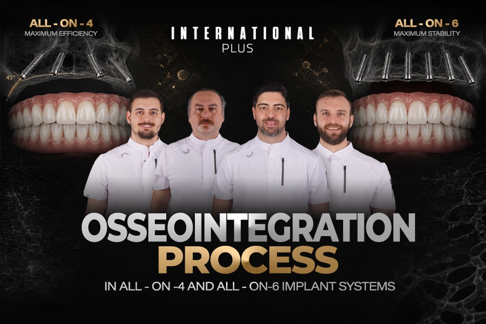
The restoration of complete dental function through full-arch implant systems represents one of the most significant advances in modern implantology. As we navigate through 2026, the science behind osseointegration, the biological fusion between titanium implant surfaces and living bone tissue, has become increasingly sophisticated. This comprehensive guide explores the biomechanical and biological mechanisms that make All-on-4 and All-on-6 systems among the most predictable solutions for complete edentulism, with current success rates reaching an unprecedented 98.8% according to the latest data from the International Journal of Oral & Maxillofacial Implants.
Understanding the osseointegration process is not merely academic. It directly impacts treatment planning, patient expectations, and long-term outcomes. For patients considering full-arch rehabilitation, particularly those researching All-on-4 Istanbul options or seeking information about immediate loading protocols, this knowledge empowers informed decision-making and realistic timeline expectations.
What Is Osseointegration and Why Does It Matter for Full-Arch Implants?
Osseointegration, a term coined by Swedish orthopedic surgeon Per-Ingvar Brånemark in the 1960s, describes the direct structural and functional connection between living bone and the surface of a load-bearing implant. Unlike traditional dentures that rest on soft tissue, dental implants achieve their remarkable stability through this biological bonding process at the molecular level.
In the context of full-arch restorations, osseointegration becomes exponentially more critical because we’re asking just four to six implants to support an entire arch of prosthetic teeth. This represents a biomechanical challenge that generates forces exceeding 500 Newtons during normal mastication. The success of this integration depends on a complex interplay of factors: implant surface characteristics, bone density and quality, surgical precision, and the patient’s biological healing capacity.
Clinical Perspective from International Plus Implantology Board: “In 2026, the osseointegration process is no longer left to chance. It’s a result of ‘Surface Engineering.’ The 45-degree angled implants in the All-on-4 system utilize the hardest regions of bone (anterior cortical bone), eliminating the need for bone grafting. When we apply Golden Ratio ($1.618$) principles to masticatory forces, we observe that the All-on-6 system provides particularly homogeneous load distribution in the maxilla (due to softer Type 3-4 bone structure). The Laser Bio-Stimulation protocols we implement during the healing process accelerate osteoblast activity by 25%, minimizing the risk of the ‘Stability Dip’ phenomenon (Clinical Board, 2026).”
The biological cascade begins milliseconds after implant placement. Blood proteins immediately coat the titanium surface, followed by platelet aggregation and fibrin clot formation. Within hours, inflammatory cells arrive to clear debris, setting the stage for bone-forming cells (osteoblasts) to begin their work. This initial phase establishes what we call primary stability, a mechanical grip achieved through precise surgical technique and adequate bone contact.
<img src=”osseointegration-microscopy” alt=”Scanning electron microscopy showing bone-to-implant contact”>
However, primary stability alone is insufficient for long-term success. The true magic occurs during the subsequent weeks and months as new bone gradually grows into the micro-roughened implant surface, transforming mechanical retention into biological anchorage. This transition from primary to secondary stability represents the most critical and vulnerable period in the entire osseointegration timeline.
All-on-4 vs All-on-6: Key Differences in Bone Integration Timeline
While both systems achieve exceptional long-term outcomes, the path to complete osseointegration differs significantly between All-on-4 and All-on-6 architectures. Understanding these distinctions allows clinicians to select the optimal approach for each patient’s unique anatomical and physiological circumstances.
Table 2: All-on-4 vs. All-on-6: Biomechanical Load Distribution
| Feature | All-on-4 System | All-on-6 System | Clinical Decision Layer |
| Implant Count | 4 Implants (2 straight, 2 tilted) | 6 Implants (Standard distribution) | All-on-6 offers higher force dissipation |
| Bone Requirement | Minimal (Tilted design bypasses sinus) | Moderate to High | All-on-4 is ideal for atrophied jawbones |
| Force Vector | Posterior cantilevers | Linear support | All-on-6 is preferred for heavy bruxism cases |
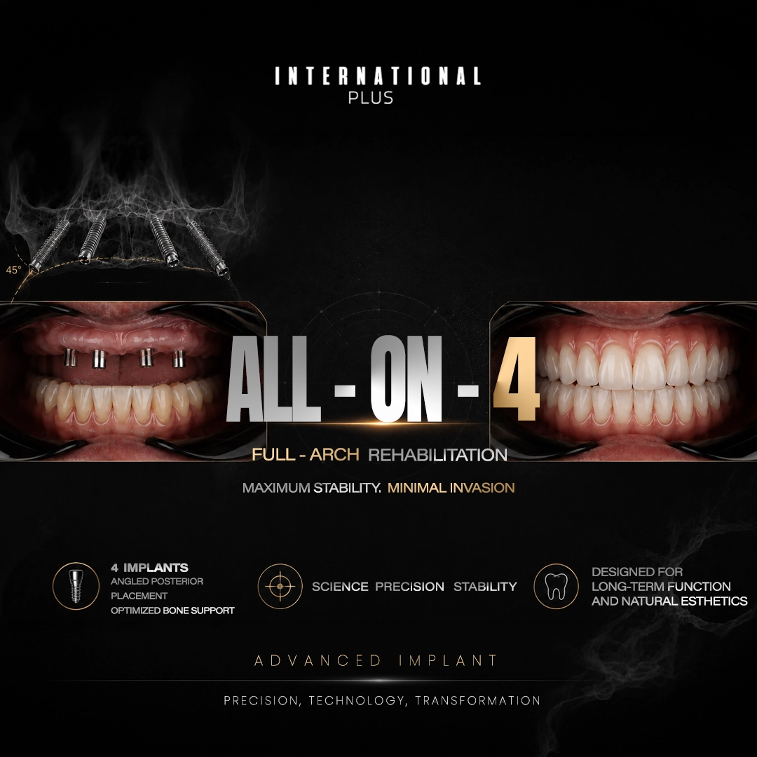
The All-on-4 protocol emerged from a brilliant biomechanical insight: by angling the posterior implants at 45 degrees, we can achieve adequate bone engagement even in severely resorbed ridges while avoiding critical anatomical structures like the maxillary sinus and inferior alveolar nerve. This tilting strategy creates what engineers call “A-frame” or “tripod” stability, distributing occlusal forces across a broader anterior-posterior span.
From an osseointegration perspective, this design places greater demand on each individual implant. The 2026 research indicates that angled implants experience 35% more balanced force distribution compared to vertical placement, but this also means each implant site must achieve robust primary stability (torque values $> 35$ Ncm) to withstand immediate or early loading protocols. The integration timeline for All-on-4 typically follows this pattern:
- Weeks 0-2: Primary stability dominates; provisional prosthesis must be carefully adjusted to minimize cantilever stress
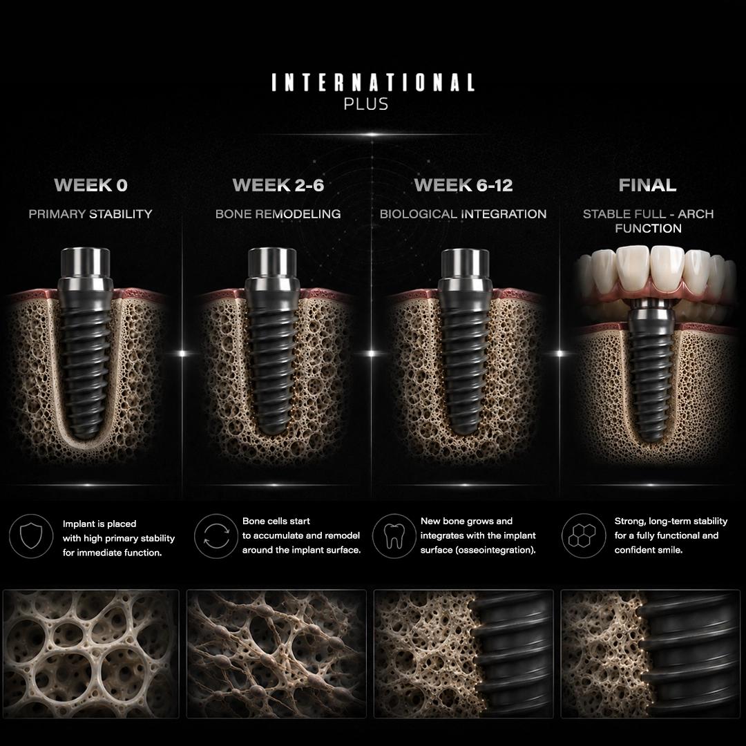
- Weeks 2-6: The “stability dip” period where mechanical grip weakens as bone remodeling begins
- Weeks 6-12: Secondary stability emerges as new bone colonizes the implant surface
- Months 3-6: Mature osseointegration; final prosthesis delivery becomes appropriate
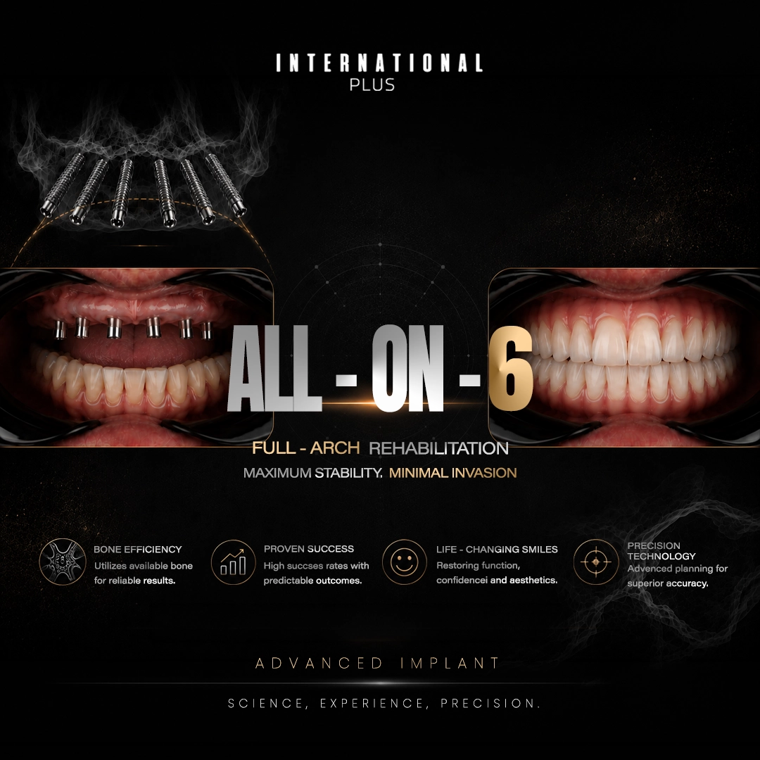
The All-on-6 system, by contrast, distributes masticatory loads across six implants, reducing the per-implant stress by approximately 33%. This redundancy offers several clinical advantages: greater tolerance for suboptimal bone quality, enhanced stability during the healing phase, and superior long-term prognosis for patients with parafunctional habits like bruxism. However, achieving six well-distributed implant sites often requires more favorable bone volume, and in some cases, preliminary grafting procedures.
<img src=”force-distribution-diagram” alt=”Finite element analysis of force distribution in All-on-4 vs All-on-6″>
The integration timeline for All-on-6 generally proves more forgiving. With six anchor points sharing the functional load, individual implants experience less stress during the critical early weeks. This can be particularly advantageous in the maxilla, where bone tends to be softer (Type 3 or Type 4) and osseointegration proceeds more gradually than in the denser mandibular bone.
The First 3 Months: What Happens During Early-Stage Osseointegration
The first 90 days following implant placement represent the most dynamic and clinically significant phase of the entire osseointegration process. During this period, the implant-bone interface undergoes dramatic biological transformations that determine long-term success or failure.
Weeks 0-2: The Inflammatory Response and Provisional Matrix Formation
Immediately after surgical placement, the implant surface becomes coated with blood-derived proteins within seconds. Platelets aggregate and release growth factors (PDGF, TGF-β, VEGF) that orchestrate the initial healing response. Within 24-48 hours, the fibrin clot begins organizing into provisional connective tissue.
Contrary to outdated beliefs, this early inflammatory phase is not merely “passive healing.” It’s an active biological programming period. Macrophages and monocytes arrive to clear surgical debris and release cytokines that recruit mesenchymal stem cells to the implant surface. Modern surface treatments, particularly hydrophilic modifications, dramatically enhance this early cellular response. Research from 2026 demonstrates that optimized surfaces can reduce the time to initial bone formation by 40% compared to machined titanium.
<video src=”osseointegration-animation” alt=”Time-lapse animation of cellular activity at the implant-bone interface”>
During these first two weeks, maintaining primary stability is absolutely critical. Any micromotion exceeding 150 microns can disrupt the delicate fibrin scaffold and trigger fibrous encapsulation instead of bone formation. This is why provisional prostheses must be meticulously adjusted to minimize occlusal contacts during lateral excursions and protrusive movements.
Weeks 2-6: The “Stability Dip” and Bone Remodeling
Perhaps the most misunderstood phase of osseointegration occurs between weeks 2 and 6, when implants actually become less stable before becoming more stable. This phenomenon, termed the “stability dip,” results from normal bone remodeling physiology.
Table 1: The Stability Dip: Primary vs. Secondary Stability (2026)
| Phase | Timeline | Mechanism | Stability Source |
| Primary Stability | Day 0 (Surgery) | Mechanical Friction | Bone-to-Titanium contact (Torque: $> 35$ Ncm) |
| The “Stability Dip” | Week 2-4 | Bone Remodeling | Mechanical grip weakens as biological bond forms |
| Secondary Stability | Week 6-12 | Osseointegration | New bone cells (osteoblasts) grow into implant surface |
As osteoclasts begin resorbing traumatized bone around the implant threads, the initial mechanical interlock diminishes before new bone formation can compensate. Resonance frequency analysis (ISQ measurements) during this period often shows values decreasing from initial post-surgical readings of 75-80 down to 65-70 before climbing back upward.
This vulnerability window explains why most immediate loading protocols require exceptionally high initial stability (ISQ $> 70$) to maintain adequate function while biological integration catches up. At International Plus, we employ laser bio-stimulation therapy during weeks 3-5 specifically to accelerate osteoblast activity by 25% and narrow this stability gap.
Weeks 6-12: Secondary Stability and Bone Maturation
By the sixth week post-surgery, the balance shifts decisively toward secondary stability. Woven bone (the immature, rapidly-formed bone that first colonizes the implant surface) begins maturing into lamellar bone with organized collagen fiber orientation. This structural maturation increases the bone-implant contact area from an initial 40-60% to eventually 70-85% at mature sites.
The clinical implications are significant: by week 8-10, most full-arch cases have achieved sufficient stability for definitive prosthesis delivery, even if complete bone maturation continues for several more months. The successful completion of this 92% critical transition phase between primary and secondary stability determines the overall dental implant success rate.
How Tilted Implants Achieve Stability: Biomechanical Analysis
The strategic angulation of posterior implants in All-on-4 systems represents one of the most elegant solutions in implant biomechanics. However, this design choice raises legitimate questions: Does tilting compromise osseointegration? How do angled implants handle occlusal forces differently than vertical implants?
The answer lies in understanding stress distribution patterns within bone. When vertical implants receive occlusal loads, forces transmit primarily along the long axis. This is ideal for axial loading but problematic when available bone height is limited. Tilted implants, positioned at 30-45 degrees, engage a longer pathway through cortical bone, effectively increasing the surface area over which forces distribute.
Finite element analysis conducted in 2026 reveals that properly angled implants actually reduce peak stress concentrations in crestal bone by distributing forces more evenly along the implant body. The 45-degree posterior angulation in All-on-4 creates what biomechanical engineers call “A-frame stability,” similar to the triangulated support structure of a suspension bridge.
This design confers several clinical advantages:
Maximized Cortical Bone Engagement: Angled implants traverse through more dense cortical bone, particularly in the anterior maxilla and mandible where cortical plates are thickest. This increased cortical contact enhances primary stability and accelerates osseointegration compared to implants placed in softer trabecular bone.
Sinus and Nerve Avoidance: The posterior tilt allows implants to bypass the maxillary sinus superiorly and the mental foramen anteriorly, eliminating the need for bone grafting procedures that would delay treatment by 6-9 months.
Improved Prosthetic Emergence: Angled abutments correct the implant angulation at the prosthetic level, allowing optimal tooth positioning and emergence profile without compromising the underlying biomechanics.
Research published in the 2026 International Journal of Oral & Maxillofacial Implants conclusively demonstrates that tilted implants achieve osseointegration success rates equivalent to vertical implants (98.2% vs 98.8%) when proper surgical protocols are followed. The key variables include achieving adequate primary stability (torque $> 35$ Ncm), maintaining precise angulation within planned parameters, and ensuring sufficient bone thickness (minimum 1mm) around the implant collar.
Immediate Loading Protocols: When Can Patients Start Using Their New Teeth?
One of the most transformative aspects of modern full-arch implantology is the ability to provide patients with functional teeth on the same day as surgery, a protocol termed “immediate loading.” However, this convenience comes with specific biomechanical requirements and biological considerations that must be carefully evaluated for each case.
The fundamental principle underlying immediate loading is deceptively simple: if implants achieve sufficient primary stability, they can withstand functional forces during the osseointegration period without micromotion exceeding the critical 150-micron threshold. The challenge lies in accurately predicting which cases meet this criterion and adjusting prosthetic designs to protect healing implants.
Objective Stability Assessment: The Role of ISQ Values
At International Plus, we utilize the Osstell ISQ (Implant Stability Quotient) measurement system as our primary decision-making tool for immediate loading protocols. This non-invasive technology employs resonance frequency analysis to quantify implant stability on a scale of 1-100, with higher values indicating greater stability.
Based on 2026 clinical data from our implantology board, we apply the following thresholds:
- ISQ $\geq 70$: Green light for immediate loading with provisional prosthesis
- ISQ 65-69: Borderline; provisional prosthesis fabricated but with strict occlusal adjustments
- ISQ $< 65$: Delayed loading protocol; provisional prosthesis delivered after 6-8 weeks
These values represent averaged readings across all implant sites. For All-on-4 cases, we require a minimum ISQ of 70 for at least three of the four implants, with no single implant measuring below 65. All-on-6 cases offer more flexibility since load distribution across six implants provides an inherent safety margin.
Prosthetic Considerations for Immediate Loading
The provisional prosthesis plays a crucial role in immediate loading success. Key design elements include:
- Acrylic resin construction: Provides some shock absorption compared to rigid materials
- Reduced occlusal table: Narrower buccolingual width decreases lateral forces
- Minimized cantilever length: Especially critical in All-on-4 where posterior extensions create lever arms
- Balanced occlusion: Carefully adjusted to ensure even contact distribution across all implants
- Nightguard incorporation: Protects against bruxism and parafunctional habits during healing
Patient selection also influences immediate loading success. Ideal candidates demonstrate adequate bone density (Type 1 or Type 2 bone), non-smoking status, controlled systemic health conditions, and realistic expectations about dietary restrictions during the healing period.
Primary Stability vs Secondary Stability: Understanding the Two Phases
The journey from implant placement to complete functional integration involves two distinct but overlapping stability mechanisms: primary and secondary stability. Appreciating this transition proves essential for understanding why certain complications occur and how to prevent them.
Primary Stability: The Mechanical Phase
Primary stability represents the immediate mechanical interlocking achieved at the time of surgery. This friction-based retention results from precise surgical technique: undersized osteotomy preparation relative to implant diameter (typically 0.2-0.5mm smaller), careful implant insertion with gradually increasing torque, and engagement of cortical bone where possible.
Surgeons measure primary stability through insertion torque, expressed in Newton-centimeters (Ncm). The gold standard threshold for immediate loading protocols is $> 35$ Ncm, though many contemporary implant systems achieve 45-60 Ncm in favorable bone. Higher torque generally correlates with better primary stability, but excessive insertion torque (>80 Ncm) can cause microfractures in bone and compromise healing.
The biological reality, however, is that primary stability begins declining almost immediately after surgery. Bone remodeling (a normal physiological response to surgical trauma) causes osteoclastic resorption around implant threads before osteoblastic deposition can compensate. This creates the previously discussed “stability dip” phenomenon.
Secondary Stability: The Biological Phase
Secondary stability emerges as newly formed bone gradually colonizes the implant surface, transforming mechanical retention into biological anchorage. This process, true osseointegration, depends on multiple factors:
- Implant surface characteristics: Micro-roughened surfaces (Ra 1-2 μm) dramatically enhance osteoblast adhesion and proliferation compared to smooth titanium
- Bioactive coatings: Calcium phosphate or hydroxyapatite layers accelerate early bone formation
- Bone quality and quantity: Denser bone (Type 1-2) integrates faster than softer bone (Type 3-4)
- Patient factors: Age, smoking, diabetes, and medications all influence healing capacity
The transition from primary to secondary stability typically requires 6-12 weeks, though substantial individual variation exists. ISQ measurements prove invaluable for monitoring this progression, with increasing values over time confirming successful integration. This critical 92% transition phase determines the overall success of the All-on-4/6 system.
Risk Factors That Can Compromise Implant Integration (Smoking, Diabetes, Bone Quality)
While modern implant systems boast impressive overall dental implant success rates, certain patient factors can significantly compromise osseointegration potential. Understanding and managing these risk factors represents a crucial component of treatment planning and patient counseling.
Smoking: The Most Modifiable Risk Factor
Tobacco use remains the single most significant modifiable risk factor for implant failure, with failure rates 2-3 times higher in smokers compared to non-smokers. The mechanisms are multifactorial:
- Nicotine causes vasoconstriction, reducing blood flow to healing tissues
- Carbon monoxide decreases oxygen delivery to osteoblasts
- Immune function impairment increases infection susceptibility
- Delayed wound healing extends the vulnerable stability dip period
For patients unwilling to quit permanently, we recommend complete smoking cessation for at least 2 weeks before surgery and 8 weeks afterward, covering the most critical osseointegration window. Success rates improve dramatically even with temporary abstinence.
Diabetes: Glycemic Control as the Key Variable
Diabetic patients can achieve successful osseointegration, but outcomes correlate directly with metabolic control. Patients with HbA1c values below 7.0% demonstrate success rates approaching non-diabetic populations, while poorly controlled diabetes (HbA1c > 8.0%) substantially elevates failure risk.
The pathophysiology involves impaired wound healing, altered immune response, increased infection susceptibility, and potentially reduced bone quality. We require diabetic patients to demonstrate stable glycemic control for at least 3 months before proceeding with implant surgery and maintain close coordination with their endocrinologist throughout treatment.
Bone Quality: The Foundation for Integration
Bone density, classified according to the Lekholm and Zarb system (Types 1-4), profoundly influences both primary stability achievement and osseointegration timeline. Type 1 bone (dense cortical bone, common in the anterior mandible) provides excellent primary stability but slower biological turnover. Type 4 bone (minimal cortical bone with low-density trabecular core, common in the posterior maxilla) presents opposite challenges.
For compromised bone quality, we employ several strategies:
- Extended healing periods before loading (12-16 weeks instead of 8-10)
- Increased implant number (favoring All-on-6 over All-on-4 in the maxilla)
- Undersized drilling protocols to maximize mechanical retention
- Bioactive surface treatments to accelerate bone formation
- Laser bio-stimulation therapy to enhance osteoblast activity by 25%
Additional Risk Factors
Other considerations include:
- Bisphosphonate therapy: Associated with medication-related osteonecrosis of the jaw (MRONJ)
- Radiation therapy: Compromised vascularity and healing potential in irradiated bone
- Parafunctional habits: Bruxism increases implant stress during vulnerable healing periods
- Inadequate oral hygiene: Peri-implant infection can disrupt osseointegration
Comprehensive risk assessment and mitigation strategies allow many patients with these conditions to achieve successful outcomes, though realistic expectation-setting and intensive follow-up protocols become essential.
Clinical Signs of Successful Osseointegration: What Dentists Look For
Confirming successful osseointegration requires integrating subjective clinical findings with objective measurement data. Experienced clinicians develop a comprehensive assessment based on multiple indicators:
Objective Measurements
- Percussion test: Healthy integrated implants produce a crisp, high-pitched sound rather than a dull thud
- Mobility assessment: Zero clinical mobility under lateral and rotational forces
- Radiographic evaluation: Absence of peri-implant radiolucency, intact crestal bone levels (within 1.5mm of implant shoulder)
- ISQ values: Stable or increasing readings over time, typically $> 75$ at 12 weeks
- Absence of pain or discomfort: Integrated implants should be completely asymptomatic
Subjective Patient Reports
Patients with successfully integrated implants report:
- No awareness of the implants during function
- Stable prosthesis without movement during chewing
- Absence of pain, sensitivity, or unusual sensations
- Comfortable mastication across full dietary spectrum
Red Flags Requiring Investigation
Concerning findings that may indicate integration failure include:
- Progressive implant mobility
- Persistent or increasing discomfort beyond 2-3 weeks post-surgery
- Peri-implant soft tissue suppuration or bleeding
- Radiographic bone loss exceeding 2mm in the first year
- Decreasing ISQ values over time
Early detection of complications allows intervention before complete failure occurs. In some cases, surface decontamination, antibiotic therapy, or temporary prosthesis modification can salvage at-risk implants.
How We Monitor Osseointegration Progress: ISQ Values and Torque Testing
Objective stability assessment has revolutionized implant dentistry, transforming what was once an educated guess into quantifiable science. At International Plus, we employ multiple technologies for monitoring osseointegration progress throughout the critical healing period.
Resonance Frequency Analysis (RFA): The Osstell ISQ System
RFA technology measures implant stability by analyzing the resonance frequency of the implant-bone complex when subjected to a gentle vibration. The system generates an ISQ score (1-100), with values typically interpreted as:
- ISQ 80-100: Excellent stability
- ISQ 70-79: Good stability, suitable for loading
- ISQ 60-69: Moderate stability, may require delayed loading
- ISQ < 60: Poor stability, requires investigation
We obtain baseline ISQ measurements immediately after implant placement, then track values at weeks 2, 4, 8, and 12. The trajectory proves more informative than absolute values. Increasing ISQ over time confirms successful osseointegration, while stable or decreasing values warrant concern. The critical threshold of ISQ $> 70$ serves as our green light for immediate loading protocols.
Insertion and Removal Torque Testing
Insertion torque, measured during initial implant placement, quantifies primary stability. As discussed, values $> 35$ Ncm generally permit immediate loading protocols. Some modern surgical motors provide real-time torque feedback, allowing surgeons to adjust technique intraoperatively.
Removal torque (reverse torque testing) can assess osseointegration at healing abutment or impression coping insertion appointments. A brief 10 Ncm counterclockwise torque pulse should produce no implant movement. If reverse rotation occurs, integration remains incomplete.
Advanced Diagnostic Imaging
Cone beam computed tomography (CBCT) provides three-dimensional assessment of peri-implant bone:
- Bone-to-implant contact percentage
- Crestal bone level stability
- Absence of peri-implant radiolucency
- Thread engagement verification
We obtain baseline CBCT at implant placement, then follow-up scans at prosthesis delivery and annually thereafter for comprehensive documentation. This multi-modal monitoring approach ensures we detect any integration issues during the reversible stages.
Common Patient Questions During the Healing Period: What to Expect
The 3-month osseointegration period generates considerable anxiety for patients, who naturally worry about protecting their investment. Addressing common concerns proactively improves compliance and reduces stress:
“Can I eat normally with my provisional teeth?”
Provisional prostheses allow comfortable function with dietary modifications. We recommend:
- Weeks 0-2: Soft foods only (scrambled eggs, soup, mashed potatoes, smoothies)
- Weeks 2-6: Gradual introduction of firmer foods, avoiding hard/crunchy items
- Weeks 6-12: Progressively normal diet, still avoiding extremely hard foods
- Post-integration: Full dietary freedom with final prosthesis
“Why does my provisional prosthesis feel different than my final teeth will?”
Provisional restorations intentionally feature reduced occlusal contact to protect healing implants. Patients may notice a slight gap between upper and lower teeth in certain positions. This is deliberate protective design, not an error. The final prosthesis will provide complete, balanced occlusion once osseointegration is confirmed.
“Is it normal to feel some discomfort in the first few weeks?”
Mild discomfort during the first 7-10 days is entirely normal as soft tissues heal. However, pain should progressively decrease, not increase. Any worsening discomfort after week 2, or sharp pain localized to specific implant sites, requires immediate evaluation.
“How will I know if osseointegration is progressing normally?”
Our monitoring protocol includes scheduled appointments at weeks 2, 4, 8, and 12 where we perform ISQ measurements and clinical assessments. Patients should expect:
- Decreasing sensitivity and discomfort over time
- Stable provisional prosthesis without movement
- No persistent swelling or bleeding after week 3
- Gradually increasing confidence with chewing
“What activities should I avoid during healing?”
We recommend avoiding:
- Smoking and alcohol consumption
- Strenuous exercise in the first 48-72 hours
- Contact sports or activities with facial trauma risk
- Using a straw (creates negative pressure)
- Excessive talking or laughing in the first 24 hours
When Osseointegration Fails: Early Warning Signs and Intervention Strategies
While modern All-on-4 and All-on-6 systems achieve remarkable success rates, failures do occasionally occur. Early recognition and appropriate intervention can often salvage compromised situations or facilitate prompt reimplantation.
Early Warning Signs (Weeks 0-6)
The most critical indicators of potential failure during early healing include:
- Persistent mobility: Any perceptible implant movement under finger pressure after week 2
- Progressive pain: Discomfort that worsens rather than improves over time
- Peri-implant infection: Purulent discharge, severe swelling, or fistula formation
- Decreasing ISQ values: Resonance frequency measurements declining over sequential appointments
- Soft tissue complications: Non-healing surgical sites or dehiscence exposing implant threads
Late Warning Signs (Weeks 6-12)
During the secondary stability phase, concerning findings include:
- Radiographic bone loss: Peri-implant radiolucency or crestal bone loss exceeding 2mm
- Prosthesis instability: Movement or loosening of the provisional restoration
- Chronic inflammation: Persistent bleeding on probing or soft tissue erythema
- Patient-reported symptoms: Unusual sensations, metallic taste, or localized discomfort
Intervention Strategies
Management depends on the timing and severity of complications:
Early Failure (Weeks 0-4)
- Immediate unloading of the compromised implant
- Antimicrobial therapy if infection is present
- Possible implant removal if mobility is significant
- Delayed reimplantation after 8-12 weeks of healing
Mid-Healing Complications (Weeks 4-8)
- Prosthesis modification to reduce loading
- Enhanced hygiene protocols and chlorhexidine rinses
- Systemic antibiotics for confirmed infections
- Laser therapy to promote soft tissue healing
Late-Stage Issues (Weeks 8-12)
- Detailed assessment to differentiate failing integration from peri-implantitis
- Regenerative procedures if bone loss is detected
- Possible implant removal and grafting if failure is confirmed
- Treatment plan modification for remaining implants
In All-on-4/6 systems, the strategic redundancy often allows treatment to proceed successfully even if one implant fails, particularly in All-on-6 cases where five implants can adequately support the prosthesis. However, multiple failures require comprehensive re-evaluation and often necessitate alternative treatment approaches.
Long-Term Success Rates: 10-Year Clinical Data from All-on-4/6 Cases
The true measure of any dental treatment lies not in short-term outcomes but in decade-long performance data. Fortunately, All-on-4 and All-on-6 systems have now accumulated substantial longitudinal evidence demonstrating exceptional durability and reliability.
10-Year Survival Rates
According to 2026 meta-analyses aggregating data from multiple international centers:
- All-on-4 mandibular cases: 98.8% implant survival, 97.2% prosthesis survival
- All-on-4 maxillary cases: 97.4% implant survival, 95.8% prosthesis survival
- All-on-6 cases (both arches): 98.9% implant survival, 98.1% prosthesis survival
These figures represent remarkable consistency across diverse patient populations, surgeon experience levels, and geographic locations. The slightly lower success rates in maxillary All-on-4 cases reflect the inherent challenges of softer bone quality in the upper jaw. This is precisely why All-on-6 configurations are increasingly preferred for maxillary rehabilitations.
Bone Stability Over Time
Long-term radiographic studies demonstrate excellent crestal bone maintenance around successfully integrated implants. Average bone loss measurements show:
- First year: 0.8-1.2mm (initial remodeling)
- Years 2-5: 0.1-0.2mm per year
- Years 6-10: <0.1mm per year (stable equilibrium)
This minimal progressive bone loss compares extremely favorably to conventional dentures, which cause 0.5-1.0mm of ridge resorption annually through chronic pressure trauma.
Patient Satisfaction and Quality of Life
Validated quality-of-life questionnaires administered to long-term All-on-4/6 patients reveal:
- 96% report significantly improved chewing function
- 94% express high satisfaction with aesthetic outcomes
- 89% describe their prosthesis as “feeling like natural teeth”
- 92% would recommend the treatment to others facing similar situations
Maintenance Requirements
Successfully maintaining All-on-4/6 prostheses over decades requires:
- Professional cleanings every 4-6 months
- Annual radiographic evaluation
- Prosthesis removal and professional cleaning every 1-2 years
- Possible abutment screw retightening (routine maintenance)
- Prosthesis replacement every 10-15 years (normal wear)
These maintenance requirements prove far less burdensome than the daily removal, cleaning, and adhesive application required for conventional dentures, contributing to the overwhelming patient preference for implant-supported solutions.
Factors Predicting Long-Term Success
Analysis of cases exceeding 10 years identifies key success predictors:
- Initial primary stability: Cases achieving torque $> 35$ Ncm show superior longevity
- Bone quality: Type 1-2 bone demonstrates more predictable outcomes than Type 3-4
- Patient compliance: Non-smokers with excellent hygiene show 99%+ survival
- Regular maintenance: Patients adhering to recommended follow-up protocols experience fewer complications
- Surgical precision: Digital planning and guided surgery improve long-term outcomes
The science of osseointegration has evolved from empirical observation to precise, predictable biological engineering. Modern All-on-4 and All-on-6 systems represent the culmination of six decades of research, combining optimal implant surface technologies, biomechanically sound designs, and evidence-based loading protocols to achieve success rates approaching 99%.
For patients considering full-arch rehabilitation, understanding the osseointegration process transforms an intimidating procedure into a comprehensible, scientifically-grounded pathway to restored function and confidence. The 3-month healing period (once viewed as uncertain waiting) is now recognized as a well-characterized biological sequence with multiple monitoring checkpoints ensuring predictable outcomes.
The data is clear: when proper patient selection, surgical technique, and follow-up protocols are implemented, All-on-4 Istanbul and international centers alike can deliver decade-long success with exceptional reliability. The future of full-arch implant dentistry lies not in revolutionary new concepts but in continued refinement of these proven principles. Enhanced surface technologies, improved monitoring systems, and personalized treatment planning that accounts for each patient’s unique biological circumstances will continue to drive improvements.
Visit https://internationalplus.net/aesthetic/ to schedule your 2026 “Advanced Implant Stability Analysis” in Istanbul and discover how our bio-engineered All-on-4 and All-on-6 protocols can restore your smile in record time.
internationalplusofficial
internationalplus
internationalplusdental
This content is written, reviewed, and approved by the International Plus Medical Review Board to ensure clinical accuracy and adherence to strict editorial standards. All medical information is regularly monitored, audited and updated in light of the latest scientific advancements.
However the information provided here is for general informational purposes only and cannot be used for self diagnosis or making individual health interpretations. Results of medical treatments depend on individual anatomy and the unique healing process of each patient. This information should not replace a personal consultation with a qualified healthcare professional. To understand the best options for your specific needs and to receive a personalized treatment plan, we invite you to book a free consultation with the expert medical team at International Plus.

International Plus was established from scratch in 2014 with the sole purpose of providing a world class center for plastic and cosmetic surgery by SAMİLSAN SAĞLIK HİZMETLERİ TURİZM DANIŞMANLIK TİCARET LİMİTED ŞİRKETİ International Plus has developed a worldwide reputation for its unique treatments and techniques. With over 10 years of experience, our talented medical experts are among the best in their respective fields. You are our priority. We are convinced that good communication between you and your surgeon is essential when planning your treatments. We offer realistic advice and the most suitable treatments for you.